Cranial Cavities & Orbit Anatomy
Orbit Anatomy
A comprehensive anatomical and educational guide to the human orbit, exploring its skeletal boundaries, communicating canals, and protective mechanisms for the visual system.
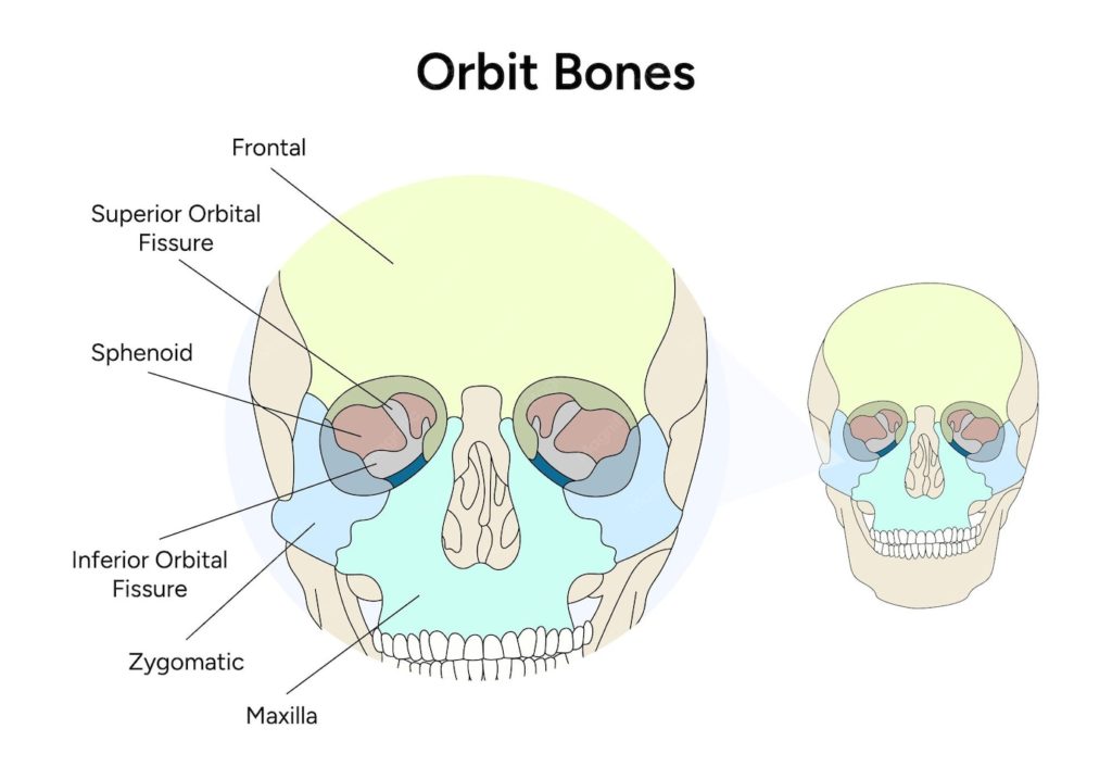
The eye socket, known anatomically as the orbit, is a paired, bilateral pyramidal skeletal cavity situated within the facial skeleton. Its base opens anteriorly onto the face, while its apex points posteromedially toward the cranial cavity. The orbit's primary biological function is to house, cushion, and protect the eyeball (globe), along with extraocular muscles, orbital fat, cranial nerves, and vascular structures. Bony Framework (The 7 Contributing Bones): Each orbital cavity is framed by the precise convergence of seven individual cranial and facial bones: Frontal bone: Forms the superior wall (orbital roof) alongside the lesser wing of the sphenoid bone. Maxilla and Zygomatic bones: Form the sturdy floor and inferolateral margin, absorbing impact and supporting ocular weight. Ethmoid, Lacrimal, and Sphenoid bones: Build the paper-thin medial wall (notably the lamina papyracea of the ethmoid) and the deep lateral boundaries. Palatine bone: Contributes a small orbital process to the posterior floor of the cavity. Openings and Communication Channels: The orbit features critical foramina and fissures that connect the orbital contents to the interior of the skull: Optic canal: Transmits the optic nerve (cranial nerve II) and the ophthalmic artery directly into the middle cranial fossa. Superior orbital fissure: A prominent cleft between the wings of the sphenoid bone that transmits cranial nerves III (oculomotor), IV (trochlear), VI (abducens), and branches of the ophthalmic nerve (V1). Inferior orbital fissure: Allows communication with the pterygopalatine and infratemporal fossae for neurovascular passage.