Thoracic Cage Osteology & Axial Skeleton
The ribs
A comprehensive anatomical guide to the human ribs, detailing their structural classification, costal groove neurovascular bundles, articulations, and respiratory biomechanics.
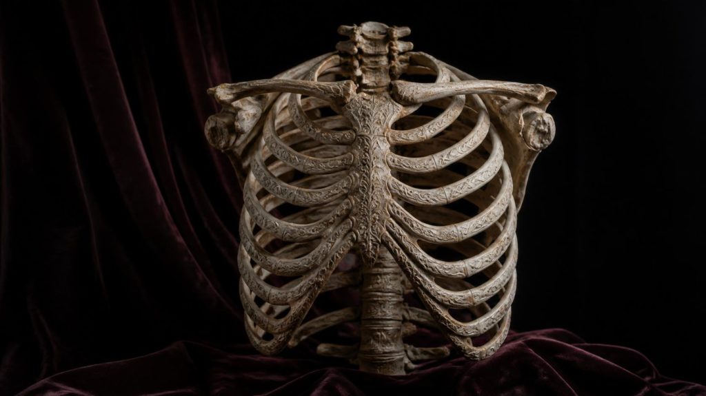
The ribs (Latin: costae) are twelve pairs of curved, flat bones that form the lateral walls of the thoracic cage. Extending from the thoracic vertebrae posteriorly toward the sternum anteriorly, they create a protective, semi-rigid framework that shields vital organs within the thoracic cavity (the heart, lungs, and great vessels) while providing structural anchor points for respiratory musculature. Classification by Anterior Attachment: The 12 pairs of ribs are categorized into three distinct functional groups: True Ribs (Vertebrosternal, Pairs 1–7): Attach directly to the sternum through their own individual costal cartilages, forming stable, protective arches over the superior thoracic viscera. False Ribs (Vertebrochondral, Pairs 8–10): Connect indirectly to the sternum; their individual cartilages merge into the costal cartilage of the rib immediately above them, creating the unified inferior costal margin. Floating Ribs (Vertebral, Pairs 11–12): Possess no anterior skeletal attachment to the sternum; their cartilaginous tips terminate freely within the abdominal wall musculature. Anatomy of a Typical Rib (Ribs 3–9): A typical rib exhibits distinct morphological landmarks along its curve: Head (Caput): Possesses two articular facets separated by a crest to articulate with the bodies of two adjacent thoracic vertebrae at the costovertebral joint. Neck (Collum): A constricted region between the head and the tubercle. Tubercle (Tuberculum): Features an articular facet that meets the transverse process of the corresponding thoracic vertebra at the costotransverse joint. Shaft (Corpus): The thin, curved body of the rib. Near the posterior angle, the curvature sharpens. Along the internal inferior margin lies the costal groove (sulcus costae), which shelters the intercostal vein, artery, and nerve (VAN sequence from superior to inferior). Respiratory Dynamics and Biomechanics: During inspiration, ribs perform two characteristic mechanical motions: the superior ribs elevate primarily in a sagittal plane ("pump-handle movement," increasing anterior-posterior thoracic diameter), whereas the lower ribs swing laterally in a coronal plane ("bucket-handle movement," increasing transverse thoracic diameter). This dynamic expansion reduces intrathoracic pressure, enabling passive pulmonary airflow.